A breast biopsy can help determine whether a suspicious spot on a breast cancer screening or painful lump needs treatment. We had an experienced physician walk us through what to expect from the procedure.
Posted
by Featured Provider Pil (Peter) Kang on Friday, August 26, 2022
Could it be cancer? If you receive a callback after a routine breast cancer screening, or begin to experience symptoms that cause concern, the looming possibility of disease can be stressful and scary. Thankfully, a minimally-invasive outpatient breast biopsy can help you get the answers you need–quickly.
“We're there to do a diagnostic evaluation and either rule out a cause or find what’s causing the issue,” says Peter Kang, MD, MBA, a radiologist at The Iowa Clinic who regularly performs the procedure.
What happens when there’s cause for concern?
Candidates for breast biopsy fall into two broad categories: Patients who are experiencing physical symptoms with an abnormal imaging finding, and patients whose routine screening shows a possible sign of disease.
“Typically, they either feel a lump themselves, or their physicians feel a lump,” Dr. Kang says. “Or they might have breast pain, or abnormal nipple discharge.”
The other population of prospective biopsy patients aren’t experiencing symptoms. However, if the radiologist performing their annual mammogram happens to see an abnormality, they may determine that it merits further evaluation.
Regardless of whether the procedure is prompted by symptoms or a screening, Dr. Kang says he engages patients in a conversation about what’s next.
“A lot of the times we talk with patients about our degree of suspicion,” Dr. Kang says. “Some masses are very suspicious—a ‘cancer until proven otherwise’ type of scenario.”
Even if the spot on a mammogram is most likely benign, because there is always a small overlap of cancer and non-cancerous masses in terms of imaging findings, Dr. Kang says many patients appreciate the reassurance of biopsy results to help reach a more certain diagnosis.
“In our clinic, when we’ve had that discussion and the patient's onboard, we typically schedule either within the same week or the following week for another encounter where we are performing a biopsy,” Dr. Kang says.
Prioritizing patient comfort and ease of biopsy.
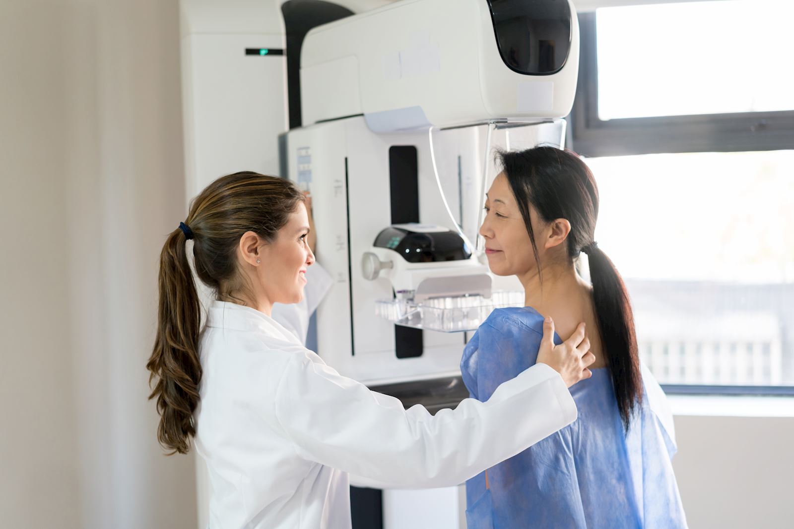
Although there are several types of breast biopsy procedures, Dr. Kang says he and his colleagues most frequently choose an ultrasound-guided method, which is the simplest and most comfortable. The goal is to use a mammogram and ultrasound image that correlate together to see that area of concern alongside what’s happening in real-time.
Dr. Kang says patients receive a local anesthetic (lidocaine, which blocks signals at the nerve ending of the skin but doesn’t cause unconsciousness) to prevent any sharp pain at the site of a small incision. Patients should expect to experience tugging sensation during the procedure, though, which happens while you are lying on your back.
“With an ultrasound probe, I can look at the mass and the needle in real-time and make sure that the needle is in the mass,” Dr. Kang says. “I can take samples and can also look for any potential complications in real-time. If there's any bleeding from the mass that's forming a hematoma, I can see them readily with an ultrasound and deal with that.”
If the finding on a mammogram (such as a calcification or architectural distortion) doesn’t correlate with an ultrasound, the physician will use the mammographic images to guide a needle to a target area. In this stereotactic or tomosynthesis guided biopsy method, you would be seated in a special mammographic chair while your breast is in compression. Occasionally, a physician will select a third method—an MRI guided biopsy—to sample the area of concern, if ultrasound or mammography aren’t correlating.
Regardless of biopsy method, the goal is to take several core solid tissue samples throughout the area of concern, which are then evaluated in a pathology lab.
Before the incision is closed—and typically, it’s such a small wound it won’t even require stiches— the medical imaging specialist will leave a tiny titanium clip, surrounded by gel foam or a similar material, to mark where the biopsy took place. The physician will confirm clip positioning and deployment with a mammogram and compress the area to ensure bleeding has stopped.
“The marker facilitates two things,” Dr. Kang says. “First, if the biopsy is benign, then in future mammograms, we don't have to guess about whether a mass has been dealt with. The other function is if we recommend a surgical excision, or if it's cancer, then the marker helps us to locate the right spot to remove the tissue.”
Healing and waiting for your results.
Following the procedure, patients typically experience a little soreness at the site. But you shouldn’t have to wait long for results. Typically, the pathology results come back within around 24 hours. So, if you have a biopsy in late morning, your results should come back by the following afternoon.
Then, Dr. Kang will call the patient with the biopsy results, to check-in on healing and outline next steps. Less than two percent of patients experience any complications (which might be an infection of hematoma); most are just anxious to have a diagnosis.
Your biopsy results should fall into one of three categories: benign, cancer, or discordant.
- A benign (noncancerous) biopsy diagnosis might be most common in a younger patient who is experiencing a fibroadenoma, a solid breast lump that is not cancer. This happens most often between the ages of 15-35 but can be found at any age in a person who has periods. This kind of diagnosis means a patient is cleared to follow a regular screening schedule from that point on.
- Discordant biopsy results occur when benign pathology results don’t match up with concerning imaging findings. “In some cases, where I was expecting cancer, but I got benign tissue, it doesn't make any sense from an imaging perspective,” Dr. Kang says. Instead of taking these results at face-value, patients with discordant findings might be referred for a surgical excision, or an MRI to further investigate the suspicious area.
- For patients who receive a cancer diagnosis, they may require a breast MRI to evaluate for extended disease. They’ll also get connected with an oncology patient navigator who will take care of treatment referrals.
Passing to the cancer ‘quarterback’.
If the mass is malignant, the breast biopsy will kick-off a multidisciplinary tumor board discussion.
“We will have in our radiology pathology, surgery, medical oncology, folks in the genetic referral center all get together to talk about the patient’s findings,” Dr. Kang says, citing the surgeon as the ‘quarterback’ of the oncology team.
“The surgeons and the medical oncologist discuss the appropriate treatment and come to a consensus on what needs to happen for a given patient,” Dr. Kang says.
Because early detection is key for breast cancer survival, scheduling regular screenings and quick follow-up can put you on track for treatment. Schedule an appointment today for a mammogram.
Meet This Featured Provider

Learn More About:
Medical Imaging & Radiology,
Women's Center
Pil (Peter) Kang, MD, MBA joined The Iowa Clinic to be part of a physician owned organization, where everyone’s part of a team. Previously he worked for a small practice in Southeast Iowa for nearly ten years and is excited for his family (and himself) to be involved in everything that Des ... Read More
Accepting New Patients
Tags
- medical imaging
- radiology
- women's center